
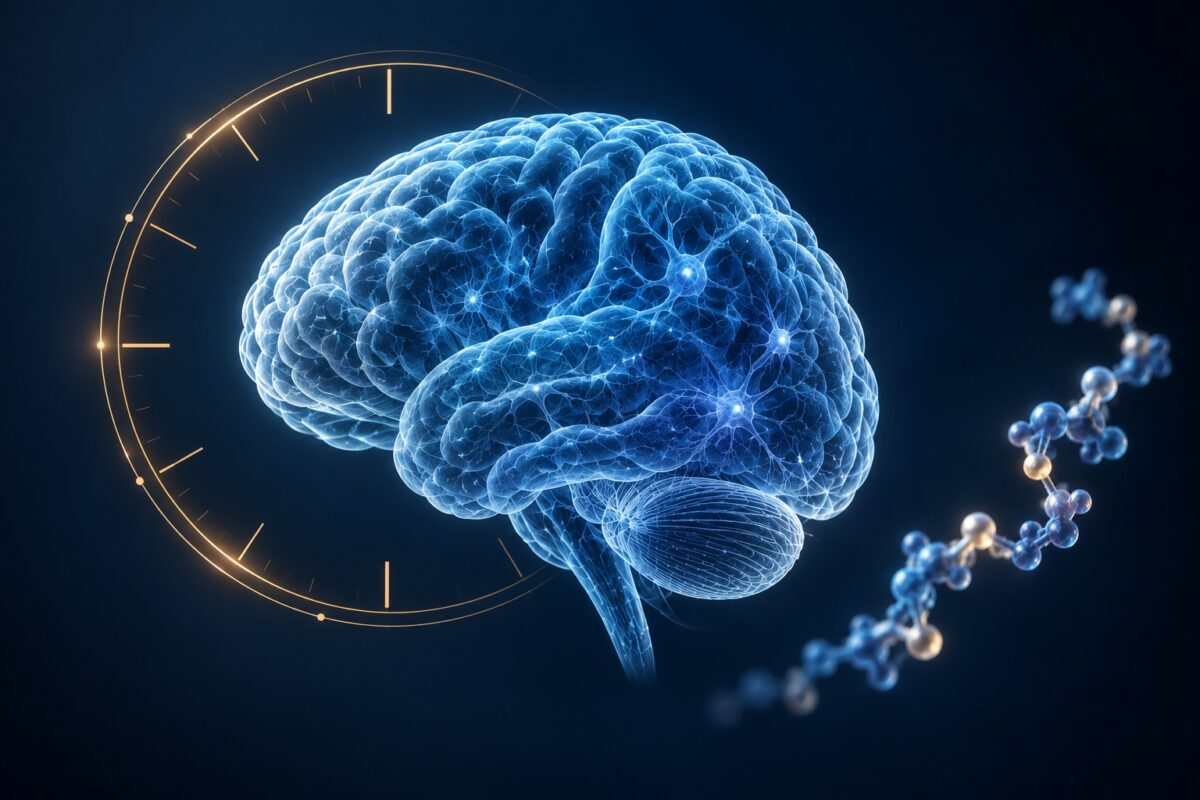
New Study: Brain-Aging Clocks Reveal How Cells Age, and How Peptides May Be Involved

What if different cells in the same brain do not show the same molecular age? A new preprint found that computer models called “brain-aging clocks” could estimate how old different types of brain cells appear, detect faster-aging signals linked to Alzheimer’s and Parkinson’s disease, and register a younger-age signal after a genetic SIRT6 experiment in mouse liver. The analysis also pointed to VGF, a gene used to make a larger protein that is cut into several bioactive peptides, suggesting a new place to look for clues about how cellular aging works.
Note: This article is intended for general information and educational purposes. It summarizes scientific research in accessible language for a broad audience and is not an official scientific press release.
Different parts of the brain do not necessarily show the same molecular signs of aging at the same time. Neurons, immune cells, and the cells that support and protect nerve fibers can follow different aging patterns. But when scientists analyze a whole piece of brain tissue at once, those differences can become blurred together.
That is where aging clocks come in. They are not actual clocks. They are computer models trained to recognize molecular patterns that tend to change as people get older. By reading those patterns, a model can estimate how old a biological sample appears. Researchers can then compare that estimate with the person’s actual age.
The new study takes that idea a step further by asking whether different types of brain cells can be “read” separately.
The study, Cell type-resolved chromatin accessibility clocks for brain aging, was posted on August 25, 2026, on the bioRxiv preprint server and has not yet undergone peer review. The authors are Patrick Z. Yu, from the Paul G. Allen School of Computer Science and Engineering at the University of Washington; Doudou Yu, from the Department of Genome Sciences at the University of Washington; Yao Xue, from the Departments of Ophthalmology and Visual Science at Yale University School of Medicine; and William Stafford Noble, affiliated with both the Paul G. Allen School of Computer Science and Engineering and the Department of Genome Sciences at the University of Washington.
What Did the Researchers Want to Find Out?
Many existing aging clocks look at chemical marks on DNA. This study focused instead on chromatin accessibility.
Chromatin is the material made of DNA and proteins that packages DNA inside the cell. Some parts of it are more “open,” meaning the cell can more easily access those DNA regions and use them to regulate genes, while other parts are more closed. These patterns can change with age and differ between cell types.
The researchers wanted to know whether these patterns could reveal how old specific types of brain cells appear at the molecular level.
They also asked whether clocks trained only on healthy human brain tissue could still recognize aging-related signals in another brain region, in other species, in Alzheimer’s and Parkinson’s disease, and after an experimental genetic change associated with younger molecular-age signals.
How Was the Study Conducted?
The team started with data from 357 healthy human donors between 15 and 100 years old. The dataset covered around 1.5 million cells from the prefrontal cortex, a region involved in complex functions such as planning and decision-making, and included more than 521,000 regions of accessible chromatin.
The cells were divided into six major groups:
- excitatory neurons;
- inhibitory neurons;
- astrocytes;
- microglia;
- oligodendrocytes;
- oligodendrocyte precursor cells, or OPCs.
Microglia are immune cells found in the brain. Oligodendrocytes produce myelin, the insulating material around many nerve fibers, while OPCs are cells that can develop into oligodendrocytes.
The researchers trained computer models to find combinations of open DNA regions that were most useful for predicting age. They created one clock using all cells together and separate clocks for each major cell type.
They then challenged the clocks with completely different datasets, including the human hippocampus, mouse hypothalamus, mouse and zebrafish retina, mouse liver, and postmortem brain samples from people with Alzheimer’s and Parkinson’s disease.
What Makes This Study Different?
Most tissue-level measurements average thousands or millions of cells together. That can hide the fact that one cell type may show a strong aging signal while another does not.
The new clocks were designed to preserve those differences.
In practical terms, instead of asking only “How old does this piece of brain tissue look?”, researchers could ask “Which kinds of cells are showing the strongest signs of aging?”
The authors note that previous work had produced a chromatin-accessibility aging clock from blood, but that a comparable cell-type-level clock had not previously been established for the brain.
Key Findings
1. The clocks estimated age across different brain-cell types
The models estimated age across all six major brain-cell groups, with excitatory neurons producing the strongest predictions. They also recognized age-related patterns in other brain regions and animal datasets, although performance generally weakened as the tissue or species became less similar to the human prefrontal cortex used for training.
2. SIRT6 overexpression was linked to a younger predicted-age signal in mice
In a separate experiment using 28 male mice, animals genetically engineered to produce more SIRT6 received younger age estimates than ordinary mice. The difference was statistically significant in the older animals.
This was a mouse-liver experiment, not evidence that SIRT6 slows human brain aging.
3. Brain immune cells showed the strongest disease-linked aging signal
The clocks were also tested on postmortem brain samples from 43 people with Alzheimer’s disease and 101 with Parkinson’s disease. Microglia, the brain’s resident immune cells, showed the strongest relationship with disease-related age acceleration among the major cell types.
The researchers also observed possible sex-related differences in severe Alzheimer’s disease, but the groups were small.
Why This Study Could Be Useful
According to the authors, the value of these clocks is not simply predicting age. They can also show which cell types and molecular pathways contribute to an aging signal, helping researchers compare healthy aging with disease or experimental interventions.
For now, this is a research tool, not a clinical test.
Where Peptides Enter the Picture
One of the most interesting clues from the aging clocks involved VGF, a gene that helps produce several small signaling molecules called peptides.
Why is that important? The researchers found a VGF-linked DNA region among the signals used to predict age in both the prefrontal cortex and the hippocampus. Another type of aging clock pointed to almost the same DNA region, which makes the VGF finding more interesting.
VGF can be processed into several bioactive peptides, including TLQP-21. The authors highlight earlier mouse research in which TLQP-21 was linked to less amyloid plaque and fewer Alzheimer’s-related changes in microglia and astrocytes.
So peptides are relevant here because the aging-clock analysis independently pointed to a gene that produces them. The study does not prove that these peptides slow brain aging, but it identifies VGF and its peptide pathway as a specific biological lead worth investigating further.
What Do the Authors Conclude?
The authors conclude that these cell-specific aging clocks can capture both normal aging and changes associated with disease or experimental interventions.
They suggest that the approach could help researchers study which cellular and molecular processes are involved when aging patterns shift.
Important Limitations
The study is a bioRxiv preprint and has not yet undergone peer review.
The clocks are research tools, not diagnostic tests. They cannot predict whether a person will develop Alzheimer’s or Parkinson’s disease, and the findings do not show that changing SIRT6 or any of the molecular pathways identified in the study will slow aging or treat disease.
The cell groups were broad, comparisons between species are imperfect, and the molecular signals found by the models show associations rather than cause and effect.
Conclusion
This study offers researchers a more detailed way to examine brain aging by looking at major cell types separately rather than relying on one average signal.
It also highlights specific biological clues, including microglia, SIRT6-related changes, and VGF-related peptide biology, that can now be investigated more closely.
The findings do not identify a treatment, but they may help researchers narrow down which cells and molecular pathways deserve further study.
The information in this article is provided for informational purposes only and is not medical advice. For medical advice, please consult your doctor.
Reference
Patrick Z. Yu, Doudou Yu, Yao Xue, William Stafford Noble. (2026). Cell type-resolved chromatin accessibility clocks for brain aging. bioRxiv 2026.08.22.746439. https://doi.org/10.64898/2026.08.22.746439