
A Picture of (Brain) Health: Powerful Brain Scans and Assessment Batteries We Use to Understand Our Most Complex Organ
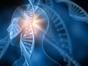
The human brain is an incredibly complex feat of nature. Capable of creating complex social structures, languages, culture, art, and science. Our brains allow us to explore and understand the universe better than any other animal on the planet ever has. But even with all of this knowledge, we are only just beginning to understand the human brain itself.
Scientists, biologists, and medical professionals are on a neverending quest to learn about the brain, and thanks to innovative brain scan technologies, we are closer than ever to unlocking the mysteries of how the brain works.
But why is it so difficult to understand how our brains function?
Brain Anatomy
The human brain is made up of billions of neurons, or brain cells, each connected in a web of synapses so dense there are more connections in a single human brain than there are stars in the observable universe.
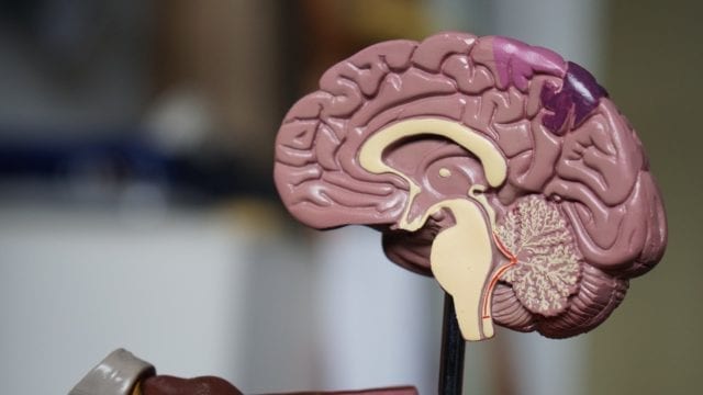
If we zoom out a little and take a holistic view of the brain, we see that the neurons are grouped into three main parts: the brainstem, the cerebellum, and the cerebrum. Each of these parts plays a unique role in how our brains function and how we think, act, and perceive the world.
The Human Brain, in Three Parts:
- Brainstem – The brainstem is located at the bottom of the brain and connects the brain and the spinal cord. Many of the automatic tasks our body performs–such as breathing, heart rate, digestion, vomiting, and more–are controlled by the brain stem.
- Cerebellum – The cerebellum is located near the bottom of the brain as well, behind the brain stem. This region of the brain is responsible for coordinating sensory input–such as what we hear, see, and smell–with our muscle movements so that we are able to understand our location within our surroundings and are able to maintain balance and posture.
- Cerebrum – The cerebrum is the largest part of the brain, covered in greyish wrinkles and folds, and is what we typically think of when we think of a ‘brain.’ Tasked with many of our higher-level brain functions, the cerebrum is responsible for interpreting what we see, hear, and gather from our various senses, as well as learning, reasoning, speaking, and emotion. Many of our fine motor movements, such as the movements required to play a musical instrument, are also controlled by this region of the brain.
Major Zones of the Cerebrum:
Each of the hemispheres of the Cerebrum is further divided into four distinct zones called lobes.
- Frontal Lobe – The frontal lobe is found on the top, forwardmost part of the brain. Many of our executive functions, such as planning, organizing, and problem-solving, are linked to this region. The frontal lobe also plays a role in short-term memory, creativity, and critical thinking.
- Parietal Lobe – The parietal lobe, found on the top of the brain, behind the frontal lobe, is responsible for helping us interpret sensory information such as taste, touch, and temperature.
- Occipital Lobe – The occipital lobe, found near the back of the brain, helps us to interpret visual information from our eyes and combine this information with past memories and experiences.
- Temporal Lobe – The temporal lobe, which can be found on the side of the brain under the frontal and parietal lobes, helps us process smells, tastes, and sound information. This part of the brain is also involved in the storage of memories.
What Tools Do We Use To Understand the Human Brain?
Though we still have a long way to go to unlock all the secrets of the human brain, new technologies, methods, and tools such as brain scans allow us to understand more about the human brain than ever before.
Brain Scans & Imaging Tools:
- PET Scan – Positron emission tomography (PET) scans are used to show which parts of the brain are active at a given moment. By injecting a tracer substance into the brain and detecting radioactive isotopes in the tracer, we can see what parts of the brain are actively using glucose, a sign of brain activity. As a specific brain region becomes active, it fills with blood, which delivers oxygen and glucose, providing fuel for that region. These areas become visible in the PET scan, thanks to the tracer substance, and allow us to create images of which areas of the brain are active during a given activity. The PET scan can only locate generalized brain areas, not specific clusters of neurons. In addition, PET scans are considered invasive and costly to perform.
- CT Scan – Computed tomography (CT) scans are used to create images of the brain by recording the levels of X-ray absorption. Subjects lay on a flat table, which is connected to a large cylindrical tube-shaped apparatus. Inside the tube is a ring that holds an X-ray emitter. As the X-ray emitter moves along the tube, sensors on the opposite side of the ring detect the amount of X-rays that pass through. Since different materials–such as skin, bone, water, or air–absorb X-rays at different rates, the CT scan can create a rough map of the features of the brain.
- MRI Scan – Magnetic resonance imaging (MRI) and functional magnetic resonance imaging (fMRI) scans are imaging tools used widely in the field of psychology. Using a strong magnetic field, MRIs create alignment within the nuclei of atoms within the tissues of the body and brain. By measuring the changes as the nuclei return to their base states, the MRI is able to create a picture of the brain’s structure. As a non-invasive procedure, with little risk to health, MRI scans can be performed on a broad range of subjects, including infants, the elderly, or pregnant mothers. Because of this, they can also be used multiple times on a single individual to map changes over time. The main difference between MRI and fMRI is that while basic MRI scans are used to image the structure of the brain, fMRI are used to map our the activity within the brain structures.
- EEG Scan – Electroencephalography (EEG) allows us to measure brain activity by placing electrodes on the scalp of a subject which sense electrical activity. EEG scans are non-invasive and allow researchers to record changes in brain activity down to the millisecond, making it one of the best options for understanding changes in the brain as they occur.
- MEG Scan – Magnetoencephalography (MEG) is a method of imaging the electrical activity in the brain through the use of magnetic fields. Extremely sensitive devices known as SQUIDs capture the activity in the brain, allowing researchers, doctors, or other professionals to understand which areas of the brain are responsible for various brain functions, or to determine the location of a pathology.
- NIRS Scan – Near-infrared spectroscopy is a brain imaging technique that uses infrared light to measure oxygen levels in the brain. By shooting infrared light through the skull and measuring the light on the other side, NIRS scans can detect brain activity in a non-invasive, though indirect, way.
Other Tools & Methods:
Though we have new tools and technology to aid us in understanding the human brain, that doesn’t mean that brain scans are the only tools we have at our disposal. Some of the best methods for understanding our brains don’t require any medical equipment at all.

- Interviews – When a patient suffers brain damage, doctors and psychologists will often perform interviews with the subject to understand how the damage to the brain affects behavior, memory, senses, or other aspects of our mental capacity. Since we already know what areas of the brain are affected by brain damage, any changes in mental ability, personality, or other brain functions may be good areas to perform additional research.
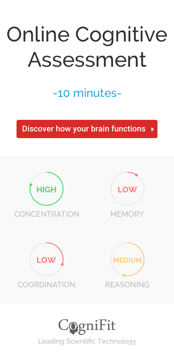
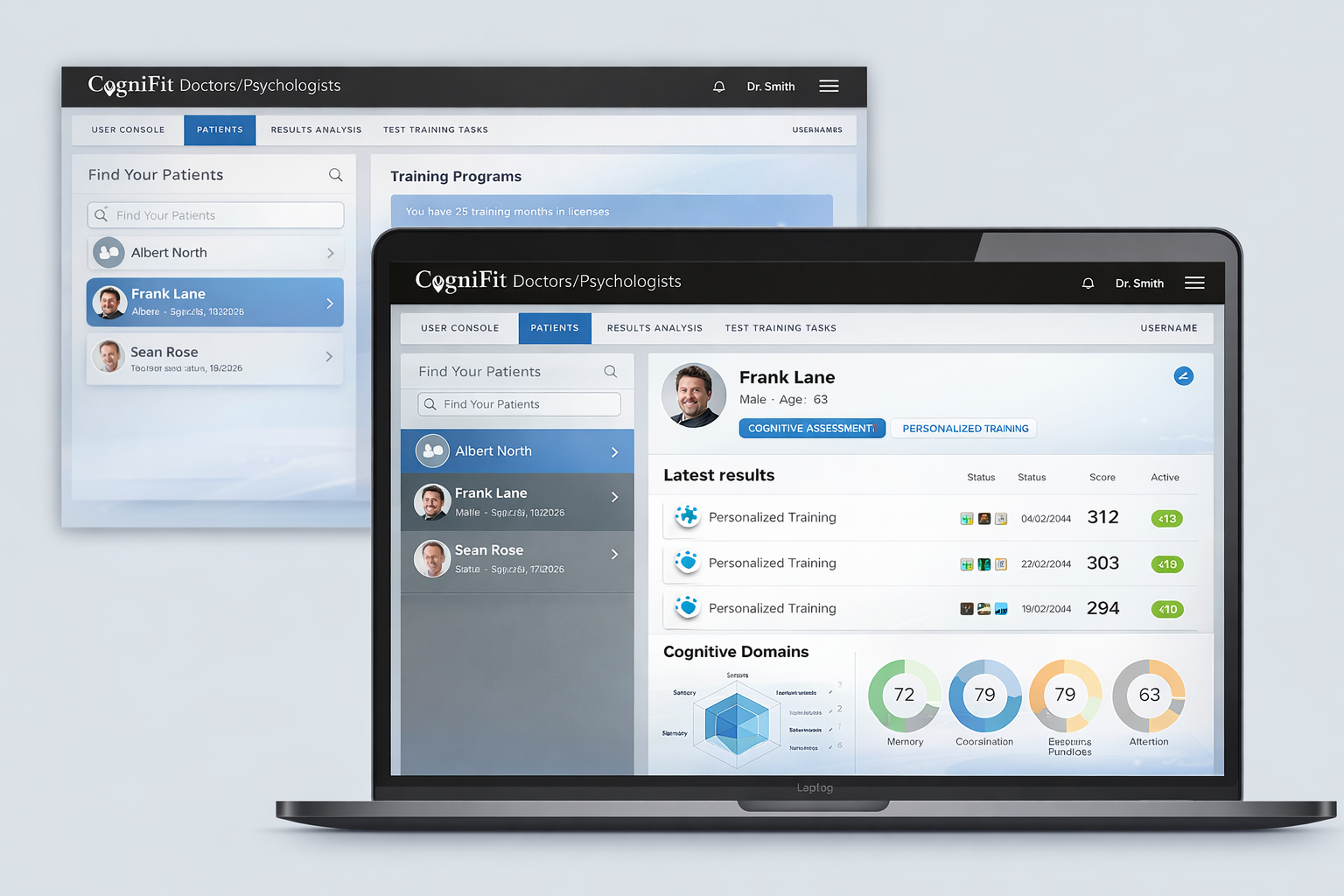
- Assessments – One of the best ways to study brain development or functioning is to have subjects complete tests or assessments. There are many assessments available for a variety of brain functions. Some of the most significant advantages to these types of evaluations are their low cost, the fact that they can be administered in nearly any setting (so you don’t have to go to a research lab or hospital) and they can be performed multiple times with no adverse effects on the health of participants. Because of this, many researchers use assessments to record changes in brain function across years of study.
Conclusion
As we continue to unlock new mysteries of the brain and create more and more powerful tools for exploring the human mind, we will continue to grow our ability to treat patients and improve the lives of people all over the world. Brain scans allow us to peak into one of the most complex systems we have ever seen. Still, it is essential to remember that it is the tool that gives us the answers, but the researchers and medical professional who interpret the results.