The Definitive Guide to the Human Brain
THE ANATOMY OF THE HUMAN BRAIN
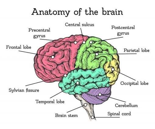
The brain is a powerful and vital organ that is essential to being alive. With that said, it would not hurt to have knowledge of the main parts of the brain and their functions. Basically, the brain has 3 parts: the cerebrum, the cerebellum, and the brain stem. Each of these parts provides different functions for the brain, and we cannot survive without them.
The Cerebrum:
Also known as the cortex, the Cerebrum is by far the largest portion of the brain and weighs about two pounds. For the record, the entire brain weighs three pounds. The cerebrum is home to billions and billions of neurons. These neurons control virtually everything we do. It controls our movements, thoughts and even our senses. Since the cerebrum has so many functions, if it’s damaged, there are many different consequences.
The cerebrum consists of four different lobes that control all of our movements. The four lobes include: the frontal lobe, parietal lobe, temporal lobe, and the occipital lobe.
The Frontal Lobe
The biggest lobe in the cortex. It is located in the front, right behind the forehead. It extends from the anterior to the central sulcus. It is the control center of your brain. The frontal lobe is involved in planning, reasoning, problem solving, judgement, and impulse control, as well as in the regulation of emotions, like empathy, generosity, and behavior. It is linked to executive functions.
The Parietal Lobe
It’s located between the central sulcus and the parietal-occipital sulcus. This part of the brain helps to process pain and tactile sensation. It is also involved in cognition.
The Temporal Lobe
It is separated from the frontal and parietal lobe by the lateral sulcus and the limits of the Occipital lobe. It is used in auditory and language processing and is also used in memory functions and managing emotions.
The Occipital Lobe
It is delimited by the posterior limits of the parietal and temporal lobes. It is involved in visual sensation and processing. It processes and interprets everything that we see. The Occipital lobe analyzes aspects like shape, color, and movement to interpret and make conclusions about visual images.
Finally, the cerebrum consists of two layers: the cerebral cortex, which controls our coordination and personality, and the white matter of the brain, which allows the brain to communicate.
The Cerebral Cortex
A thin layer of gray matter that grooves around itself, forming a type of protuberance, called convolutions, that give the characteristic wrinkled look to the brain. The convolutions are delimited by grooves or cerebral sulci and those that are especially are deep are called fissures.
The cortex is divided into two hemispheres, right and left, and they are separated by the interhemispheric fissure and joined by a structure called the corpus callosum which allows transmission between the two. Each hemisphere controls a side of the body, but this control is inversed: the left hemisphere controls the right side, and the right hemisphere controls the left side. This phenomenon is called brain lateralization.
White Matter
White matter is the subway of the brain. It connects the different parts of gray matter in the cerebrum to another. Like a subway/metro, this type of matter remains underneath it all (the surface in life, gray matter in the brain) and this underneath part is filled with different passages, links, and paths to take- each one with a different destination and purpose.
It’s known to be white because this type of matter is myelin rich. Myelin is a fatty-rich substance that causes the matter to appear white. In reality, the matter is a pinkish-white. In adults, the matter is about 1.7-3.6% blood and takes up about 60% of the brain!
The Limbic System:
Your limbic system functions range from regulating your emotions to storing your memories to even helping you to learn new information. Your limbic system is one of the most essential parts of the brain that help you live your daily life. The primary structures that work together in your limbic system are the amygdala, the hippocampus, the thalamus and hypothalamus, the cingulate gyrus, and the basal ganglia. All these parts help you to be active in society, engage in social relationships, and be a well-rounded person. To learn more about the interesting ways your limbic system impacts your life, sit back and get in-tuned with all of its hard-working employees!
The Amygdala
Shaped like a small almond, the amygdala is located in each of the left and right temporal lobes. It’s known as “the emotional center of the brain,” because it is involved in evaluating the emotional intake of different situations or emotional intelligence (for example, when you feel happy because you received an awesome grade on your math exam or when you might be frustrated because the heavy traffic is making you late for work).
The amygdala is what makes the brain recognize potential threats (like if you are hiking in the lone woods and suddenly you hear the loud footsteps of a bear coming toward you). It helps your body prepare for fight-or-flight reactions by increasing your heart and breathing rate. The amygdala is also responsible for understanding rewards or punishments, a psychological concept known as reinforcement coined by the classical and operant conditioning experiments of Ivan Pavlov.
The Hippocampus
The Hippocampus is a small subcortical seahorse shaped structure that plays an especially important role in the formation of memory, both in classification and long-term memory. Among its main functions are the mental processes related to memory consolidation and the learning process. As well as processes associated with the regulation and production of emotional states and spatial perception.
The Thalamus
It is similar to the re-transmission station of the brain: it transmits the majority of perceived sensory information (auditory, visual, and tactile), and allows them to be processed in other parts of the brain. It is also used in motor control.
The Hypothalamus
It is a gland located in the center area of the base of the brain that has an especially important role in the regulation of emotions and many other corporal functions like appetite, thirst, and sleep. The functions of the Hypothalamus are essential to our daily life. It is responsible for maintaining the body’s systems, including body temperature, body weight, sleep, mating, levels of aggression and even emotional regulation. Most of these functions are regulated by a chain of hormones that inhibit or release between themselves.
The Cingulate Gyrus
This part is located in the middle of your brain next to the corpus callosum. Not much is known about the cingulate gyrus, but researchers suggest that this is the area that links smell and sight with pleasurable memories of previous experiences and emotions because it provides a pathway from the thalamus to the hippocampus. This area is involved with your emotional reaction to pain and how well you regulate aggressive behavior.
The Basal Ganglia
This area is an entire system within itself located deep in the frontal lobes. It organizes motor behavior by controlling your physical movements and inhibiting your potential movements until it gets the instructions to carry them out, based on the circumstances that you are in. The basal ganglia also participate in rule-based habit learning; choosing from a list of potential actions; stopping yourself from undesired movements and permitting acceptable ones; sequencing; motor planning; prediction of future movements; working memory; and attention. It is made up of a few structures, such as:
The Caudate Nucleus
The caudate nucleus sends messages to your frontal lobe, specifically to your orbital cortex (just above the eyes) which alerts you that something is not quite right with the physical situation you are in (usually during tense or anxious moments), so you should take action to fix your uneasiness.
The Putamen
The putamen lies directly underneath the caudate and controls your coordinated automatic behaviors, like riding a bike, driving a car, working on an assembly line, and any other task that doesn’t really involve upper-level thinking.
The Nucleus Accumbens
The nucleus accumbens is a brain part involved in functions such as motivation, reward, or positive behavioral reinforcement. The role of nucleus accumbens is to integrate motivation along with the motor action. Its function is to transfer relevant motivational information to the motor cells in order to obtain a certain reward or satisfaction. An imbalance is related to many psychiatric and neurological disorders such as depression, obsessive-compulsive disorder, bipolar disorder, anxiety disorders, Parkinson’s disease, Huntington’s disorder, obesity and drug abuse.
The Cerebellum:
From Latin, meaning “little brain,” the cerebellum is a two-hemisphere structure located just below the rear part of the cerebrum, right behind the brain stem. Representing about 11 percent of the brain’s weight, it is a deeply folded and highly organized structure containing more neurons than all of the rest of the brain put together. The surface area of the entire cerebellum is about the same as that of one of the cerebral hemispheres.
The cerebellum is the second largest part of the brain, and it plays a significant role for our motor skills. It is located at the base of the brain, and damage to it can lead to decline in your motor skills. Besides motor control, the cerebellum has other different functions. One function that it has is to maintain our balance and posture. Another major function of the cerebellum is that it helps control the timing and force of various muscles.
Motor learning is another function of the cerebellum, and it has the biggest impact on skills that require trial and error. Even though it is mostly associated with motor control, the cerebellum has some control of our cognitive functions, such as language.
The Brain Stem:
Even though the brainstem is small, it controls many important functions in our bodies. Some functions of the brainstem include breathing, arousal, awareness, blood pressure, heart rate and digestion. It also controls our sleep patterns, body temperature, heart rhythms and even our hunger and thirst. In addition, it regulates the central nervous system.
The brain stem is the oldest and deepest area of the brain. It is often referred to as the reptilian brain because it resembles the entire brain of a reptile. The brainstem is also the smallest part of the brain and sits beneath your cerebrum in front of your cerebellum—and it connects the cerebrum to the spinal cord. Parts of the brainstem include: the midbrain, medulla oblongata and the pons.
The Midbrain
It is the structure that joins the posterior and anterior brain, driving motor and sensory impulses. Its proper functioning is a pre-requisite for the conscious experience. Damages to this part of the brain are responsible for some movement problems, like tremors, stiffness, strange movements, etc.
The Medulla Oblongata
It helps control our automatic functions, like breathing, blood pressure, heart rate, digestion, etc.
The Pons
The Pons, also known as the Annular Protuberance, is the portion of the base of the encephalon that is located between the medulla oblongata and midbrain. It connects the spinal cord and the medulla oblongata to the superior structures in the hemispheres of the cerebral cortex and/or the cerebellum. It is used in controlling the brain’s automatic functions and it has an important role in the awake-state levels and consciousness and sleep regulation.
The Spinal Cord:
The Spinal Cord is a long, whitish cord that is located in the vertebral canal and connects the encephalon to the rest of the body. It acts as a type of information highway between the encephalon and the body, transmitting all of the information provided by the brain to the rest of the body.
THE CENTRAL NERVOUS SYSTEM: NERVES, NEURONS, & NEUROTRANSMITTERS
Have you ever stopped to think about how the Nervous System works? How is your body organized? How does it really work? What structures make up the Nervous System? We are full of tracks that come and go loaded with data, electrical currents, chemicals, etc. at different rates and for different purposes.
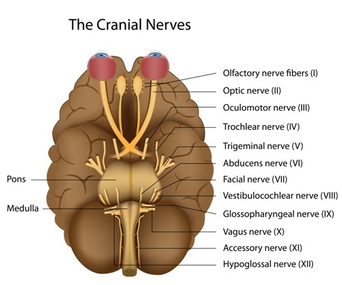
Cranial Nerves:
12 pairs of cranial nerves enable us to perform our daily routine in a comfortable and efficient way, as they take part of the information of our senses to the brain and the brain to some of our muscles and viscera. Here is a small guide to know a little more about what are the cranial nerves, their anatomy, their classification, and their function.
As shown in the image above, the 12 pairs of cranial nerves have an associated Roman numeral. These numbers range from 1 to 12 corresponding in each case to the pair in question.
Each cranial nerve has a specific function. The next image shows how this person’s head is portrayed through numbers according to the cranial nerve functions. Would you dare to say what function each cranial pair has according to its number in the drawing?
Before starting, it’s important to point out the order that this explanation will have will be according to the corresponding Roman number assigned to the cranial nerve.
The Olfactory Nerve (I)
It’s the first of the 12 pairs of cranial nerves. It’s a sensory nerve, in charge of transmitting olfactory stimuli from the nose to the brain. Its actual origin is given by the cells of the olfactory bulb. It is the shortest cranial pair of all.
The Optic Nerve (II)
This cranial pair is the second of the 12 pairs of cranial nerves and it is responsible for conducting visual stimuli from the eye to the brain. It is made of axons from the ganglion cells of the retina, that take the information of the photoreceptors to the brain, where later it will be integrated and interpreted. It emerges in the diencephalon.
The Oculomotor Nerve (III)
This cranial nerve is also known as the common ocular motor nerve. It is the third of the 12 pairs of cranial nerves. It controls eye movement and is also responsible for pupil size. It originates in the midbrain.
The Trochlear Nerve (IV)
This nerve has a motor and somatic functions that are connected to the superior oblique muscle of the eye, being able to make the eyeballs move and rotate. Its nucleus also originates in the mesencephalon as well as the oculomotor nerve. It is the fourth of the 12 pairs of cranial nerves.
The Trigeminal Nerve (V)
It is a mixed cranial nerve (sensitive, sensory and motor), being the largest of all cranial nerves, it is the fifth of the 12 pairs of cranial nerves. Its function is to carry sensitive information to the face, to convey information for the chewing process. The sensory fibers convey sensations of touch, pain, and temperature from the front of the head including the mouth and also from the meninges.
The Abducent Nerve (VI)
It is also known as the external ocular motor cranial nerve and it is the sixth of the 12 pairs of cranial nerves. It is a cranial motor pair, responsible for transmitting the motor stimuli to the external rectus muscle of the eye and therefore allowing the eye to move to the opposite side from where we have the nose.
The Facial or Intermediate Nerves (VII)
This is another mixed cranial pair since it consists of several nerve fibers that perform different functions, like ordering the muscles of the face to create facial expressions and also send signals to the salivary and lacrimal glands. On the other hand, it collects taste information through the tongue. It is the seventh of the 12 pairs of cranial nerves.
The Vestibulo-Cochlear Nerve (VIII)
It is a sensory cranial nerve. It is also known as the auditory and vestibular nerve, thus forming vestibulocochlear. He is responsible for balance and orientation in space and auditory function. It is the eighth of the 12 pairs of cranial nerves.
The Glossopharyngeal Nerve (IX)
It is a nerve whose influence lies in the tongue and pharynx. It collects information from the taste buds (tongue) and sensory information from the pharynx. It leads orders to the salivary gland and various neck muscles that help with swallowing. It also monitors blood pressure. It is the ninth of the 12 pairs of cranial nerves.
The Vagus Nerve (X)
This nerve is also known as pneumogastric. It emerges from the medulla oblongata and supplies nerves to the pharynx, esophagus, larynx, trachea, bronchi, heart, stomach and liver. Like the previous nerve, it influences the action of swallowing but also in sending and transmitting signals to our autonomous system, to help the regulate activation and control stress levels or send signals directly to our sympathetic system. It is the tenth of the 12 pairs of cranial nerves.
The Accessory Nerve (XI)
This cranial pair is named the spinal nerve. It is a motor nerve and could be understood as one of the “purest”. It governs movements of the head and shoulders by supplying the sternocleidomastoid and trapezius muscles in the (anterior and posterior) regions of the neck. The spinal nerve also allows us to throw our heads back. Thus, we would say that it intervenes in the movements of the head and the shoulders. It is the eleventh of the 12 pairs of cranial nerves.
The Hypoglossal Nerve (XII)
It is a motor nerve which, like the vagus and glossopharyngeal, is involved in tongue muscles, swallowing and speech. It is the twelfth of the 12 pairs of cranial nerves.
What are Nerves Made From:
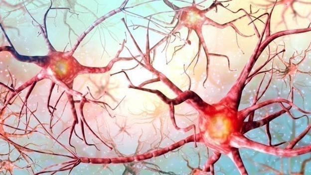
Neurons are the building blocks of the central nervous system. A neuron’s primary role is to communicate information. It communicates via electrical impulses or using specific chemicals such as neurotransmitters (what are the different types of neurotransmitters?). The neuron has 3 distinct parts. The dendrites, the cell body and the axon. Each structure plays a specific role in ensuring neurons are able to send and receive signals and connect with other neurons.
The dendrites are connected to the cell body. They conduct messages from axon of other neurons and pass the message onto the cell body. The cell body sits between the dendrites and the axon. It determines the strength of the message it receives from the dendrites. If it is strong enough, it will send the message down the axon. The axon is connected to the cell body. It conducts the message from the cell body and passes it on to other neurons.
The Dendrites
Dendrites are branch-like structures structures surrounding the cell body. They receive electrical and chemical messages from other neurons, which are collected in the cell body. These messages are either inhibitory or excitatory in nature. If the message is inhibitory, the cell body will not transmit the message to the axon. However, if the message is excitatory in nature, then the cell body will send the message down the axon and pass it to other neurons.
The Soma (or Cell Body)
Also known as the soma, the cell body is a ball-like structure. It contains the control center of the neuron, also known as the nucleus. Together, the cell body and the nucleus control the functions of the nerve cell. To be able to do this, the cell body contains organelles or really tiny organs in the nucleus.
Each organelle has a unique job. First and foremost, the most important organelle, the nucleus, regulates all cell functions. It also contains the cell’s DNA, which is essentially the neuron’s blueprint. The nucleus is another organelle that serves a vital purpose to the functioning of the neuron. It nucleolus produces ribosomes, which are essential to protein production. The cell body is also home to the endoplasmic reticulum, Golgi apparatus, and mitochondria. The mitochondria is the neuron’s fuel source, it produces all the energy needed for the nerve cell to function properly.
The endoplasmic reticulum and the Golgi apparatus, work together, with the rest of the organelles in the nucleus to produce and transport protein. The protein produced by the cell body, are the key ingredients, to build new dendrites. Building new dendrites enable the neuron to make new connections with other neurons. As well as making proteins, the cell body is also responsible for making chemicals, also known as neurotransmitters, which neurons use as signals. Neurotransmitters can serve and inhibitory or excitatory function to the neuron.
The Axon
The axon is long and slender, and it projects electrical impulses away from the cell body. The axon communicates with other neurons. When the electrical or chemical message reaches the axon terminal (end of the axon), The axon terminal release neurotransmitters into the synapse (small junction between two neurons). The neuron uses the synapse to communicate and send messages to other nerve cells.
How Nerves Communicate:
A synapse is the space between two neurons which allows for neural communication, or synaptic transmission. Synapses are found throughout the body, not just located in the brain. They project onto muscles to allow muscle contraction, as well as enable a multitude of other functions that the nervous system covers.
As a synapse is the gap in between two neurons, we need to establish which neuron sends out the signals and which neuron receives those signals.
The Presynaptic Neuron
The presynaptic neuron is the neuron that initiates the signal. At many synapses in the body, presynaptic neurons are vesicles filled with neurotransmitters. When the presynaptic neuron is excited by an action potential, the electrical signal propagates along its axon towards the axon terminal. This excitation signals the vesicles in the presynaptic neuron, filled with neurotransmitters, to fuse with the membrane of the axon terminal. This fusion allows for the neurotransmitters to be dumped into the synaptic cleft.
The Postsynaptic Neuron
The postsynaptic neuron is the neuron that receives the signal. These signals are received by the neuron’s dendrites. When there are neurotransmitters present in the synapse, they travel across the gap in order to bind to receptors on the postsynaptic neuron. When a neurotransmitter binds to a receptor on the postsynaptic neuron’s dendrite, it can trigger an action potential. That action potential can then be propagated and influence further communication.
In the nervous system there are two main types of synapses: chemical synapses and electrical synapses. Thus far, for simplicity and understanding the basics of how a synapse functions only chemical synapses have been discussed. This poses the question: why does the nervous system need two types of synapses?
Chemical Synapses
Chemical synapses are any type of synapse that uses neurotransmitters in order to conduct an impulse over the small gap in between the presynaptic and postsynaptic neurons. These types of synapses are not in physical contact with each other. Since the transmission of a signal depends on the release of chemicals, a signal can only flow in one direction. This direction is downward from presynaptic to the postsynaptic neuron.
As previously stated, these types of neurons are widely spread throughout the body. The chemicals released in these types of synapses ways excite the following neuron. The neurotransmitters can bind to the receptors on the postsynaptic neuron and have an inhibitory effect as well. When inhibition occurs, signal propagation is prevented from traveling to other neurons.
Chemical synapses are the most abundant type of synapse in the body. This is because various neurotransmitters and receptors are able to interpret signals in a large combination. For instance, a neurotransmitter and receptor combination may inhibit a signal on one postsynaptic neuron but excite a large amount of other postsynaptic neurons.
Chemical synapses allow for flexibility of signaling that makes it possible for humans to engage in high-level tasks. However, this flexibility comes at a cost. Chemical synapses have a delay due to the need for the neurotransmitter to diffuse across the synapse and bind to the postsynaptic neuron. This delay is very small but still is an important point when comparing the two types of synapses.
Electrical Synapses
Electrical synapses are types of synapses that use electricity to conduct impulses from one neuron to the other. These synapses are in direct contact with each other through gap junctions. Gap junctions are low resistance bridges that make it possible for the continuation of an action potential to travel from a presynaptic neuron to a postsynaptic neuron. Due to their physical contact, electrical synapses are able to send signals in both directions, unlike chemical synapses. Their physical contact and the use of sole electricity make it possible for electrical synapses to work extremely fast.
Transmission is also simple and efficient at electrical synapses because the signal does not need to be converted. Another key difference between chemical and electrical synapses is that electrical synapses can only be excitatory. Being excitatory means that an electrical synapse can only increase a neuron’s probability of firing an action potential. As opposed to being inhibitory, which means that it decreases a neuron’s probability of firing an action potential. This can only be done by neurotransmitters. Despite being extremely fast, these types of excitatory signals cannot be carried over great lengths.
Electrical synapses are mainly concentrated in specialized brain areas where there is a need for very fast action. The best example of this is the large amount of electrical synapses in the retina, the part of the eye that receives light. Vision and visual perception are our dominant senses, and our eyes are constantly receiving visual sensory information. This information also runs on a feedback loop when we interact with our environment, which means that we receive information from our surroundings and immediately create an appropriate response to it. This is why it makes sense that electrical synapses are seen in a large concentration here. The fast action, multiple directions, and efficiently all allow for prime functionality.
How Nerves Communicate – Neurotransmitters:
You’ve probably heard of how dopamine plays a role in feelings of pleasure, or how serotonin levels influence depression. But neurotransmitters do so much more than make us feel happy or sad. Not only do they influence our mood, but they also influence how our hearts beat, how our lungs breathe, and how our stomachs digest the food we eat.
Neurotransmitters interact with receptors on the dendrites of the neuron, much like how a lock and key work. The neurotransmitters have specific shapes that fit into a receptor that can accommodate that shape. Once the neurotransmitter and the receptor are connected, the neurotransmitter sends information to the next neuron to either fire an action potential, or to inhibit firing. If the neuron gets the signal to fire, then the whole process starts over again along the chain of neurons.
Here are some of the most important neurotransmitters:
Dopamine
Dopamine plays many different roles in the brain, depending on the location. In the frontal cortex, dopamine acts as a traffic officer by controlling the flow of information to other areas of the brain. It also plays a role in attention, problem-solving, and memory. And you’ve probably heard how dopamine plays a role in things that give us pleasure. So, if you were to eat a piece of chocolate, dopamine would be released in some areas of the brain, allowing you to feel enjoyment, motivating you to eat more chocolate.
Serotonin
Serotonin is known as an inhibitory neurotransmitter, meaning that it doesn’t give the next neuron the signal to fire. Serotonin is involved with mood, as well as your sleep cycle, pain control, and digestion. In fact, the majority of serotonin in the body can be found in the gastrointestinal tract, and only about 10% is located in the brain. Aside from aiding in digestion, serotonin can also help with forming blood clots and increasing sex drive.
Acetylcholine
Acetylcholine (ACh) plays a major role in the formation of memories, verbal and logical reasoning, and concentration. ACh has also shown to help with synaptogenesis or the production of new and healthy synapses throughout the brain. Acetylcholine comes from the chemical known as choline, which can be found in foods such as eggs, seafood, and nuts.
Acetylcholine also plays a significant role in movement. A nerve cell can release ACh into a neuromuscular junction, which is a synaptic connection between a muscle fiber and a nerve cell. When ACh is released, it causes a series of mechanical and chemical reactions that result in the contraction of muscles. When there is a lack of ACh in the neuromuscular junction, the reactions stop, and the muscle relaxes.
GABA
GABA is also an inhibitory neurotransmitter that helps to balance any neurons that might be over-firing. This inhibitory ability becomes especially helpful when it comes to anxiety or fear because the release of GABA helps to calm you down. In fact, caffeine actually works to inhibit GABA from being released, so that there is more stimulation in the brain.
GABA also plays a role in vision and motor control. Some drugs work to increase the levels of GABA in the brain. This increase helps with epilepsy and helps to treat the trembling found in patients with Huntington’s disease.
Noradrenaline (norepinephrine)
These might sound like two big and confusing words because you’ve probably heard about adrenaline (epinephrine) before. Before we go any further, let’s define these terms. Another name for adrenaline is epinephrine. Epinephrine is a hormone that is secreted by the adrenal gland, which is a gland that rests on top of the kidneys. Hormones are molecules that are released into the bloodstream. Noradrenaline is also known as norepinephrine.
Norepinephrine is a neurotransmitter, meaning that it is used for interactions between neurons. Noradrenaline is an excitatory neurotransmitter that helps to activate the sympathetic nervous system, which is responsible for your “fight or flight” response to a stressor. Norepinephrine also plays a role in attention, emotion, sleeping and dreaming, and learning. When it is released into the bloodstreams, it helps to increase heart rate, release glucose energy stores, and increase blood flow to the muscles.
BRAIN SCANS & RESEARCH
The human brain is an incredibly complex feat of nature. Capable of creating complex social structures, languages, culture, art, and science. Our brains allow us to explore and understand the universe better than any other animal on the planet ever has. But even with all of this knowledge, we are only just beginning to understand the human brain itself.
Types of Brain Scans & Imaging Tools:
Today we still do not have a clear-cut picture of the whole brain in itself. Not every network has been mapped, but we have moved forward a substantial amount. The development of non-invasive and invasive neuroimaging methods and their use for research and medical purposes was a definite breakthrough.
We have methods that can view the cortical areas of the brain. Other techniques look at cortical columns and different layers. We have methods that can record a single cell by itself. Going even further, we can look at the soma of the neuron, the dendrite and, separately the axons. We can even look at the synaptic connections between the two neurons.
Here are some of the most common types of brain imaging tools:
PET Scan
Positron emission tomography (PET) scans are used to show which parts of the brain are active at a given moment. By injecting a tracer substance into the brain and detecting radioactive isotopes in the tracer, we can see what parts of the brain are actively using glucose, a sign of brain activity. As a specific brain region becomes active, it fills with blood, which delivers oxygen and glucose, providing fuel for that region.
These areas become visible in the PET scan, thanks to the tracer substance, and allow us to create images of which areas of the brain are active during a given activity. The PET scan can only locate generalized brain areas, not specific clusters of neurons. In addition, PET scans are considered invasive and costly to perform.
CT Scan
Computed tomography (CT) scans are used to create images of the brain by recording the levels of X-ray absorption. Subjects lay on a flat table, which is connected to a large cylindrical tube-shaped apparatus. Inside the tube is a ring that holds an X-ray emitter. As the X-ray emitter moves along the tube, sensors on the opposite side of the ring detect the amount of X-rays that pass through. Since different materials–such as skin, bone, water, or air–absorb X-rays at different rates, the CT scan can create a rough map of the features of the brain.
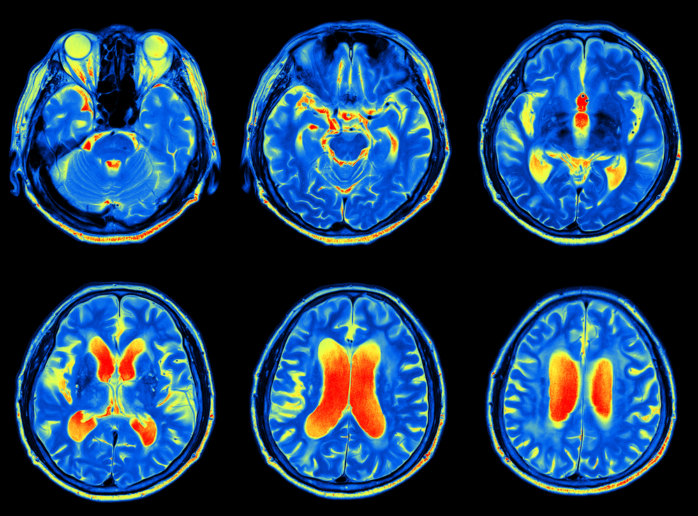
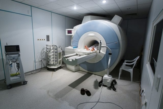
MRI Scan
Magnetic resonance imaging (MRI) and functional magnetic resonance imaging (fMRI) scans are imaging tools used widely in the field of psychology. Using a strong magnetic field, MRIs create alignment within the nuclei of atoms within the tissues of the body and brain. By measuring the changes as the nuclei return to their base states, the MRI is able to create a picture of the brain’s structure.
As a non-invasive procedure, with little risk to health, MRI scans can be performed on a broad range of subjects, including infants, the elderly, or pregnant mothers. Because of this, they can also be used multiple times on a single individual to map changes over time. The main difference between MRI and fMRI is that while basic MRI scans are used to image the structure of the brain, fMRI are used to map our the activity within the brain structures.
fMRI Scan
An upgrade from the MRI – Functional Magnetic Resonance Imaging detects the blood-oxygen-level dependent contrast imaging (BOLD) levels in the brain which are the changes in the blood flow and it not only gives the anatomical structures but the functions as well. Various colors will change depending on which part of the brain is active.
The big drawback with this technique is the fact that it does not directly measure brain activity, but BOLD signal so we cannot for sure say that the activity that we find via fMRI studies is fully accurate and is produced by neurons.
DTI Scan
Diffusion Tensor Imaging, a technique based on MRI and it measures the way the water can travel through the white matter in the brain. It can show the activity as the colored area on the image. It’s particularly good in detecting concussions so can be used in clinical applications which is a huge advantage. Again, it does not measure direct brain activity which is a huge disadvantage and sometimes it also distorts the images. DTI has a quite low spatial resolution.
EEG Scan
Electroencephalography (EEG) allows us to measure brain activity by placing electrodes on the scalp of a subject which sense electrical activity. EEG scans are non-invasive and allow researchers to record changes in brain activity down to the millisecond, making it one of the best options for understanding changes in the brain as they occur.
MEG Scan
Magnetoencephalography (MEG) is a method of imaging the electrical activity in the brain through the use of magnetic fields. Extremely sensitive devices known as SQUIDs capture the activity in the brain, allowing researchers, doctors, or other professionals to understand which areas of the brain are responsible for various brain functions, or to determine the location of a pathology.
NIRS Scan
Near-infrared spectroscopy is a brain imaging technique that uses infrared light to measure oxygen levels in the brain. By shooting infrared light through the skull and measuring the light on the other side, NIRS scans can detect brain activity in a non-invasive, though indirect, way.
TMS Scan
The electric field that TMS, or Transcranial Magnetic Stimulation, is able to generate is able to interfere with the action potentials that are happening in the brain. It’s a highly invasive technique and is able to be used in research applications for the workings of many diseases and pathologies. What we do know is that repetitive TMS is able to produce seizures so, obviously, it has some sort of side effects and needs to be used with caution.
BRAIN HEALTH & FUNCTION
Once upon a time, researchers and scientist theorized that the brain stops developing within the first few years of life. The connections the brain makes during the ‘critical period’ are fixed for life. However, there is mounting evidence, from human and animal studies, that this view underestimates the brain. The brain has a remarkable ability to continually make new connections throughout our life, it has an extraordinary ability to compensate for injury and disease by ‘rewiring’ itself. Neuroplasticity, or brain plasticity, refers to this ability to form new connections, reorganize already established neural networks and compensate for injury and disease.
Brain Plasticity:
There are many types of brain plasticity. Positive brain plasticity, which enhances healthy functioning of the brain. Negative brain plasticity, which promotes unhealthy functioning of the brain. Synaptic plasticity occurs between neurons, whereas non-synaptic plasticity occurs within the neuron. Developmental plasticity occurs during early life and is important for developing our ability to function. Injury induced plasticity is the brain’s way of adapting to trauma.
Positive Neuroplasticity
Positive brain plasticity involves changes to structures and functions of the brain, which results in beneficial outcomes. For example, improving the efficiency of neural networks responsible for higher cognitive functions such as attention, memory, mood.
There are many ways in which we can promote neuroplastic change. Positive brain plasticity is when the brain becomes more efficient and organized. For example, if we repeatedly practice our times tables, eventually, the connections between different parts of the brain become stronger. We make less errors and can recite them faster.
Cognitive Behavioral Therapy, meditation, and mindfulness can all promote brain plasticity. These practices improve neural function, strengthen connections between neurons.
Negative Brain Plasticity
Negative brain plasticity causes changes to the neural connections in the brain, which can be harmful to us. For example, negative thoughts can promote neural changes and connections associated with conditions such as depression, and anxiety. Also overuse of drugs and alcohol enhances negative plasticity by rewiring our reward system and memories.
Synaptic Plasticity
Synaptic plasticity is the basis for learning and memory. Furthermore, it also alters the number of receptors on each synapse (synapses are the connections between neurons that transmit chemical messages). When we learn new information and skills, these ‘connections’ get stronger. There are two types of synaptic plasticity, short-term and long-term. Both types can go in two different directions, enhancement/excitation, and depression. Enhancement strengthens the connection, whereas depression weakens it.
Short-term synaptic plasticity usually lasts tens of milliseconds. Short-term excitation is a result of an increased level of certain types of neurotransmitters available at the synapse. Whereas short-term depression is a result of a decreased level of neurotransmitters, long-term synaptic plasticity lasts for hours.
Long-term excitation strengthens synaptic connections, whereas long-term depression weakens these connections. As synaptic plasticity is responsible for our learning ability, information retention, forming and maintaining neural connections, when this process goes wrong, it can have negative consequences. For example, synaptic plasticity plays a key role in addiction. Drugs hi-jack the synaptic plasticity mechanisms by creating long-lasting memories of the drug experience.
Non-Synaptic Plasticity
This type of plasticity occurs away from the synapse. Non-synaptic plasticity makes changes to the way in which the structures in the axon and cell body carry out their functions. The mechanisms of this types of plasticity are not yet well understood.
Developmental Plasticity
In the first few years of life, our brains change rapidly. This is also known as developmental plasticity. Although it is most prominent during our formative years, it occurs throughout our lives. Developmental plasticity means our neural connections are constantly undergoing change in response to our childhood experiences and our environment. Our processing of sensory information informs the neural changes. Synaptogenesis, synaptic pruning, neural migration, and myelination are the main processes through which development plasticity occurs.
Synaptogenesis
Rapid expansion in formation of synapses so that the brain can successfully process the high volume of incoming sensory stimuli. This process is controlled by our genetics.
Synaptic Pruning
Reduction of synaptic connections to enable the brain to function more efficiently. Essentially, connections that aren’t used or aren’t efficient are ‘pruned’ or ‘disconnected’.
Neural Migration
this process occurs whilst we are still in the womb. Between 8 and 29 weeks of gestation, neurons ‘migrate’ to different parts of the brain.
Myelination
This process starts during fetal development and continues until adolescence. Myelination is when neurons are protected and insulated a myelin sheath. Myelination improves the transmission of messages down the neuron’s axon.
Injury-Induced Plasticity
Following injury, the brain has demonstrated the extraordinary ability to take over a given function that the damaged part of the brain was responsible for. This ability has been noted in many case studies of brain injury and brain abnormalities. Some stroke sufferers have displayed remarkable feats of recovering functions lost due to brain damage.
Neurogenesis:
You may have heard at some point in your life that you cannot grow new brain cells. You may have been taught that from the moment you are born to when you die you can only lose brain cells. It is believed that this is due to hits to the head, consuming alcohol and narcotics, and from lack of cognitive stimulation. Well do not despair because your brain is not in danger, you can in fact “grow” new brain cells in a process called neurogenesis.
Scientists at Carnegie Mellon University‘s Center for Cognitive Brain Imaging (CCBI) have used a new combination of neural imaging methods to discover exactly how the human brain adapts to injury.
When one brain area loses functionality, a “back-up” team of secondary brain parts immediately activates, replacing not only the unavailable area but also its confederates (connected areas), the research shows.
The research found that as the brain function in the Wernicke area decreased following the application of rTMS (transcranial magnetic stimulation), a “back-up” team of secondary brain areas immediately became activated and coordinated, allowing the individual’s thought process to continue with no decrease in comprehension performance.
The Brain-Body Connection:
The human brain is a marvel of evolution, capable of creating breathtaking works of art and music, developing complex systems of culture, language, and society, and uncovering mysteries of the universe through science, technology, and mathematics. But even a healthy brain couldn’t do any of these things without a healthy body to support it.
Anyone who has had to perform on stage or give a speech in front of a large group of people knows that the stress and anxiety, supposedly mental phenomenon, can manifest in physical discomforts such as “Butterflies” in our stomachs, sweaty palms, and increased heart rate.
Similarly, when we find ourselves receiving praise or affection, the feelings of happiness and euphoria we experience are readily apparent when our cheeks blush, our eyes dilate, and in extreme cases, we can even begin to cry from joy.
By taking care of our bodies, we can help to ensure our brains are functioning at their best. Although there is no single exercise or diet that is right for everyone – each person should speak to their nutrition or health professional to understand the best regimen for themselves – there are specific general rules of thumb for exercise and diet that can help just about anyone improve their brain health.
BRAIN VARIATIONS

Every person thinks and acts a little differently than the other 7 billion on the planet. Scientists now say that variations in brain connections account for much of this individuality, and they’ve narrowed it down to a few specific regions of the brain. This might help us better understand the evolution of the human brain as well as its development in individuals.
Each human brain has a unique connectome – the network of neural pathways that tie all of its parts together. Like a fingerprint, every person’s connectome is unique. Researchers found very little variation in the areas of the participants’ brains responsible for basic senses and motor skills.
The real variety arose in the parts of the brain associated with personality, like the frontoparietal lobe. This multipurpose area in the brain curates sensory data into complex thoughts, feelings or actions and allows us to interpret the things we sense.
Brain Differences Based on Gender
There are some differences found in the brains of males and females, however it’s important to note that factors influencing brain development in both males and females include, not only biology, but also the environment. We must keep in mind that culture, and social constructions have an important role in how our brains develop.
In 1989, the National Institute of Mental Health (NIMH) initiated a large-scale longitudinal study of typical brain development, which to date has acquired data regarding brain development and function from over 1000 children (including twins and siblings) scanned 1-7 times at approximately two-year intervals. This study has provided much of the information we know today about the differences between the developing male and female brain.
Studies utilizing this data have found that the peak brain size in females occurs around 10.5 years, while the peak occurs around 14.5 years in males. The other areas most frequently reported as being different are the hippocampus and amygdala, with the larger size or more rapid growth of the hippocampus is typically reported in females, and the amygdala is larger or grows more rapidly in males. The hippocampus controls emotion, memory, and the autonomic nervous system, and the amygdala is responsible for instinctual reactions including fear and aggressive behavior. Because of the larger hippocampus, girls and women tend to input or absorb more sensory and emotive information than males do.
Brain Differences Based on Handedness
The brain has two hemispheres, that each specialize to govern specific tasks. The right hemisphere of the brain controls the left side of the body and is associated with mainly spatial perception tasks, face recognition, and understanding music. The left hemisphere controls the right side of the body and is associated with more computational tasks such as math and logic. The specialization of each side of the brain is important because it allows for maximizing neural processing.
Handedness can correlate to what function each hemisphere specializes in, which allows the brain to be almost anatomically symmetrical, but functionally asymmetrical. Functional asymmetry, or lateralization, allows for each hemisphere to work in tandem when processing the world around us.
Brain Differences Based on Age
We often forget we were once teenagers ourselves. Their angst, impulsivity, and the crazy desire to live for fun makes them seem as if they are from another world. These characteristics are due to the teenage brain. The teenage brain undergoes a series of changes during cognitive development and is easily influenced by a number of factors. Physically, an adult and a teenager are near the same size.
But when it comes to the brain, there are vast differences. The teenage brain relies on the amygdala. The amygdala is reactive, stimulating a strong emotional response. When making decisions and problem solving, a teenager relies mainly on emotions. An adult’s cognitive processes are carried out using the developed prefrontal cortex—the area of the brain that causes us to think prior to behaving. Thoughts and decisions of an adult are less reactive and more logical and rational.
BRAIN & DRUGS
Consuming drugs affects the brain’s limbic system. This brain structure is in charge of awarding the satisfaction of our vital needs with a pleasant sensation or pleasure (when we are hungry and we eat, we feel pleasure). When we consume drugs, we feel a similar sensation based off of artificial pleasure, which is what leads to the start of a drug addiction.
Drugs happen to be chemical substances and they are able to affect the brain in various ways. They usually do so by interfering with how neurons communicate with one another. They can either enhance or diminish the sending, receiving and processing information functions. In the normal functioning after the neuron sends the information onto the next neuron and the neurotransmitters or chemical messengers are not needed website anymore, they are re-uptaked back or ‘cleaned’ up. Some drugs will block this re-uptake, therefore, leaving an enormous amount of these neurotransmitters in the synaptic cleft which causes the message to be enhanced and disrupts further communication. Amphetamine and cocaine do that.
Other drugs like heroin and marijuana are able to mimic a neurotransmitter by attaching themselves to the post-synaptic receptors. Therefore, they can activate other neurons but not in the same way as a neurotransmitter would. Because of that, they will send different messages along the pathways of the network, therefore, altering its normal functioning.
How Drugs Affect the Brain
When people use drugs continuously for a very long period of time their brain becomes used to this much amount of dopamine. The brain will start to compensate by naturally either making a smaller amount of dopamine and decreasing the receptors where dopamine binds in an attempt to regulate things back into homeostasis. Dopamine will therefore not be able to produce as much pleasure anymore, for any activities. That’s why it’s so difficult for a person who abuses drugs to get back into normal life – the pleasure they used to feel from regular activities diminishes.
How Cocaine Affects the Brain
Although there are many neurotransmitters, dopamine and GABA are the two altered from cocaine use. The neurotransmitter, dopamine, oversees the body’s pleasure and reward system. Cocaine acts on dopamine by signaling a sudden release of dopamine in the area between neurons (synapses) and tricking the brain’s pleasure response. The abundance of dopamine is why users feel euphoria upon exposure. Normally a second neurotransmitter known as GABA counteracts the raised dopamine levels. However, the process is unsuccessful because cocaine blocks its release. Continual use of cocaine overwhelms the nervous system. Eventually, neurons in the brain can no longer communicate when the drug induces a rush of dopamine. The dopamine receptors are damaged.
How Marijuana Affects the Brain
The endocannabinoid system is a biological system to maintain homeostasis. For the body to function properly, its conditions require balance. The heart rate must be within normal limits, temperature cannot be too hot or cold, and more. Cells in the body naturally produce endocannabinoids, which communicate with the nervous system and perform this role. Endocannabinoids attach to cannabinoid receptors on the surface of cells and are eventually destroyed by metabolic enzymes.
Marijuana, however, interferes with the endocannabinoid system. Cannabinoids from marijuana like THC bind to cannabinoid receptors, overloading the system and preventing naturally produced endocannabinoids from their regular tasks. The reward system consists of a series of brain structures from the ventral tegmental area to the hypothalamus that mediates reward. Neurons in these brain areas release dopamine upon pleasurable behaviors such as food or sex. Marijuana acts on the brain’s reward system.
As the THC attaches to cannabinoid receptors, the reward system is activated, and the user no longer responds as strongly to other pleasurable experiences. This is evidence of the addictive nature of marijuana. Scientists have taken a recent interest in how marijuana interacts with the brain’s reward system. Published in the journal, Human Brain Mapping, long-term marijuana users had more activity in the reward system on magnetic resonance imaging when shown marijuana related objects than non-users, and they had a reduction of brain stimulation when given alternative cues like their favorite fruit.
How Prescription Stimulant Use Affects the Brain
Scientists have discovered college-aged individuals who occasionally use stimulant drugs, such as cocaine, amphetamines and prescription drugs such as Adderall, display brain changes that may put them at higher risk for developing a serious addiction later in life.
A study from the University of California, San Diego School of Medicine, published in the Journal of Neuroscience, showed that occasional users have slightly faster reaction times, suggesting a tendency toward impulsivity. The most striking difference, however, occurred during the “stop” trials. Here, the occasional users made more mistakes, and their performance worsened, relative to the control group, as the task became harder. The brain images of the occasional users showed consistent patterns of diminished neuronal activity in the parts of the brain associated with anticipatory functioning and updating anticipation based on past trials.
BRAIN FACTS
The Human Brain is (relatively) BIG:
Relative to size, human brains are much bigger than other mammals. In fact, our brains are over three times bigger than mammal’s brains similar in size. As you can imagine, there is no correlations between the animals’ absolute brain sizes and cognitive abilities. Cows, for example, have larger brains than just about any species of monkey, but unless they are very, very good at hiding it, cows are almost certainly less cognitively capable than most, if not all, “lesser-brained” primates.
The Human Brain is Inverted:
The right side of the brain interacts with the left side of our bodies, and the left side of the braininteracts with the right side of our bodies. Both sides of the brain have specific functions, but sometimes the two sides of the brain interact and work together. The right brain focuses on the expression and reading of emotions, understanding metaphors, and reading faces while the left brain is far more logical, focusing on language skills, analytical time sequence processing and skilled movement.
Size Doesn’t Always Mean Power:
Having a bigger brain does not mean you are more intelligent. Clearly, there is more to intelligence than brain size, or Albert Einstein, one of the smartest people who ever lived, who had an average brain size, would have been out of luck! It is important to take into consideration how to actually define intelligence.
The Human Brain is Full of Fat:
The brain is composed nearly 60% by fat, because without it, we could not live. People who eat a diet low in omega 3 fatty acids are more likely to suffer accelerated wear and tear on the brain. The brain is regarded as the fattest organ in our entire bodies. It has the highest concentration of fat present in a single organ in a healthy human being.
The Electrical Activity Produced by The Brain Forms A Pattern of Brain Waves:
This electrical activity of the brain changes depending on the activity that is being done. For example, the brainwaves of a sleeping person are very different from the brainwaves of someone that is awake.
The Texture of The Brain Is Similar To Tofu:
Experts say our brain has a consistency similar to that of tofu or gelatin. Fatty tissues, blood vessels, and water found in the brain give it that same consistency.
The Brain Feels No Pain:
Since there are no pain receptors in the brain, it is incapable of feeling pain. This feature explains why neurosurgeons can operate on brain tissue without causing a patient discomfort, and, in some cases, can even perform surgery while the patient is awake, as we saw before.
Emotions Are Found in The Primitive Structure of Your Brain:
The limbic system is composed of a set of cerebral structures that are considered very primitive in evolutionary terms, being placed in the superior part of the brainstem, below the cortex. These structures are fundamentally involved in the development of many of our emotions and motivations, particularly those related to survival such as fear, anger, and emotions linked to sexual behavior.