
Neuroimaging: What is it and how can it map the brain?
One of the ways psychology has progressed came from the use of various neuroimaging methods. In terms of experimental psychology history, neuroimaging started with the cognitive revolution. Many scientists realized that understanding the brain plays an enormous role in the external behavior. Scientists also use neuroimaging methods and technique prevention, diagnosis and treatment for different neurological diseases.
Today we still do not have a clear cut picture of the whole brain in itself. Not every network has been mapped, but we have moved forward a substantial amount. The development of non-invasive and invasive neuroimaging methods and their use for research and medical purposes was a definite breakthrough.
Neuroimaging-What can we map?
When one thinks about the brain and the nervous system, one can think of many things to map. Of course, we have the brain itself, its parts and the functions of the anatomical functions. We have neuroimaging techniques who deal exactly with that. Despite the anatomy, however, there are many neuroimaging methods that try to look at things on a more microscopic level.
We have methods that can view the cortical areas of the brain. Other techniques look at cortical columns and different layers. We have methods that can record a single cell by itself. Going even further, we can look at the soma of the neuron, the dendrite and, separately the axons. We can even look at the synaptic connections between the two neurons.
Neuroimaging- Method Classification
Neuroimaging methods also do not just encompass the spatial resolution. We try to look into proteins, organelles, bacteria, mammalian cells, the brain of various species and, finally, human brains. Many neuroimaging methods also differ by the temporal resolution. They differ by how quickly they are able to detect an event that happens in the brain. These neuroimaging methods differ by milliseconds, seconds, minutes, hours and days. They also differ by the spatial resolution. Some methods can show anatomical structures well, while others cannot. Apart from that, the variety of the neuroimaging methods differs by how non-invasive and invasive they are.
If one can imagine, scientists use a lot more non-invasive neuroimaging methods in research. Not many regular participants agree to something that can potentially alter their brain functions. Medical practitioners are a lot more likely to use invasive neuroimaging methods in an attempt to treat certain diseases. Various patients with neurological diseases benefit on a daily basis from the invasive neurological methods. In some cases, the patients themselves are able to control the stimulating method.
Electrophysiological techniques
For many years now we know that neurons are able to generate electric potentials. We also know that the synaptic activity of the nervous matter is similar to a battery. It acts as an electric generator.
If we recall the first class in physiology we took, we can roughly remember the structure of the neuron. Words like the cell body or the soma, dendrites and an axon come to mind. Dendrites seem to be able to receive electrical signals. Axon sends electrical signals to the dendrite of the next neuron. The cell body combines the signals from the previous neurons. Then it sends another signal along the axon for the next neuron.
Within the neurons themselves, we are able to distinguish two different types of electrical activity.
1-Action potentials
The action potential is a very common concept that many students learn in their first class on the nervous system. The entire process happens for about 1 ms and culminates with the release of neurotransmitters in the end of the axons.
- The stimulus from a previous neuron activates the voltage gates on sodium channels which will cause the influx of positively charged sodium to the cell.
- This depolarizes the membrane. Sometimes the depolarization of the membrane is able to reach the threshold.
- If that happens, a series of events happen in order to send the signal along the axon to the next neuron. This is what we call an action potential.
- The potassium channels are still closed and since we have an influx of sodium, the membrane becomes more positive on the inside then it does on the outside.
- After that, the channels for sodium close and, therefore, the influx of sodium stops as well.
- That’s when the potassium channels stay open and the potassium comes out of the cell and makes the inside of the cell negative one more time. This repolarization of the neuron can lead to the overall voltage to be below the original resting potential
- This happens due to the fact that the potassium channels stay open a little longer. This ends in hyperpolarization. During this period a new action potential cannot happen and this is what we call a refractory period of the neuron.
- Scientists cannot record action potentials via surface electrodes. As of today, we are not able to record potentials from a single neuron. What we can record is the second type of electrical activity. We can, however, use intracranial electroencephalography (EEG) to measure them which happens to be an invasive technique.
2- Post-synaptic potentials
They last for hundreds of milliseconds and it is the addition of the potential from various neurons that happen at the same time. We are able to record the potentials together. Researchers can easily record these potentials from surface electrodes. Electroencephalography (EEG) can measure these types of potentials.
So, in the end, we are able to distinguish two principal types of neuroimaging methods that measure the electrical activity of the neuron.
Two principal types of electrophysiological techniques
- Single-cell recordings
- These recordings are able to measure a number of different action potentials every second. The electrodes will be place inside a single cell or nearby a neuron which makes the technique invasive.
- This technique can be useful for researchers who want to understand how single cells work.
- Due to the fact that this technique allows measuring single neurons, we are able to see how specific these cells are.
- A paper published saying that single neurons were firing to Jennifer’s Aniston’s face and nobody else’s. This level of object recognition falls under very high-level vision neurons and the paper gained a lot of attention due to such a strange working of a single neuron. (1)
- Event-related potentials (ERP)
- These recordings get the summation of different electrical potentials for a variety of neurons (millions of them). This technique places electrodes on the skull, therefore, they are surface electrodes.
Electroencephalography & Event-Related Potentials (ERP)
Since we now know that the brain produces electrical potentials, we are able to measure them. Electroencephalography helps us do that. Scientists can place various electrodes on the surface of the scalp and then measure the bio-electrical activity that the brain produces. Event-related potentials (ERP) are the potentials from various neurons that happen as a result of different stimuli given by the scientist to the participant. Stimuli and the tasks that the researchers assign can range from motor, to sensory and cognitive.
So the scientists are able to measure where and when the neurons will spike as a result of a certain assigned stimuli. Researchers have been able to find various ERP components or similarly distributed neurons that fire at the same time. They found various ERP components related to language, visual attention, auditory components (famous concepts like the mismatch negativity) and many others.
Other neuroimaging methods
Magnetoencephalography (MEG)
Neuroimaging methods don’t just stop at measuring the electrical activity of the neurons. Another famous brain imaging technique is MEG – it records magnetic fields. Electrical currents that already occur in the brain generate magnetic fields. MEG is able to directly measure the brain function which is a huge advantage when comparing it with other techniques. Apart from that, it has very high temporal resolution and high spatial resolution which is one of the rarest things when it comes to brain research. Usually, neuroimaging methods are either higher in spatial resolution or in temporal resolution, not both.
MEG is non-invasive. Scientists are able to use it with other neuroimaging methods at the same time – like EEG. One big disadvantage of MEG comes from the fact that in order to get the magnetic fields, a special room that gets rid of other types of magnetic interference needs to be built. Due to this, the machine is quite costly, but one of the best methods for measuring brain activity as of today.
Other famous types of brain imaging do not measure direct brain activity, however, they have quite good spatial resolution and are often used for clinical and diagnostic purposes.
Positron Emission Tomography (PET)
This technique gives an image of brain activity, however, in order to produce that image radioactive material needs to be either inhaled or injected by the participant. The image will then be produced due to this radioactive material going to the areas of the brain that are active.
Computed Tomography Scan (CT Scan)
This technique is able to produce brain images as well. It is able to show the anatomy of the brain, however, not the functions themselves which are a serious drawback especially if we consider the fact that X-ray lights need to go through the head to produce the image.
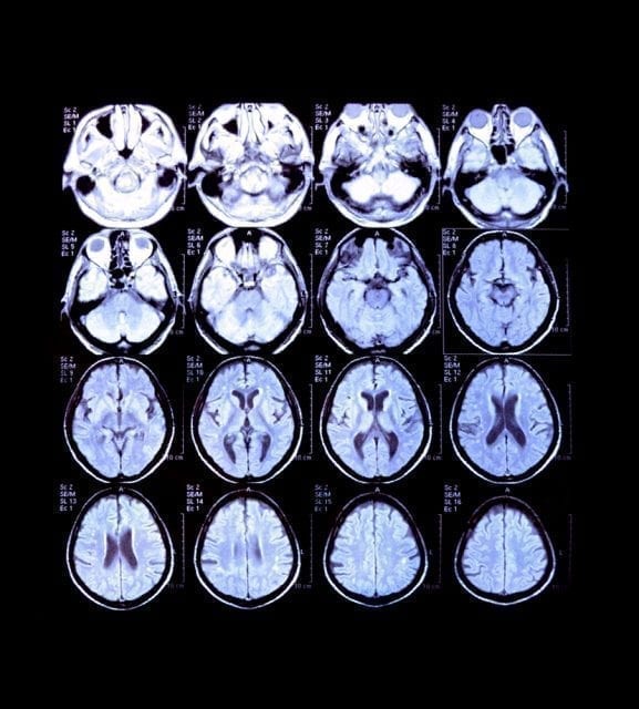
Magnetic Resonance Imaging (MRI)

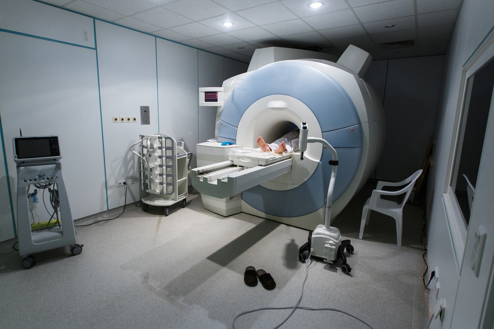
One of the most common techniques nowadays. It gives an image of anatomical structures in the brain. It is non-invasive, but the patient must remain still in the MRI chamber which could prove to be quite painful for those suffering from claustrophobia. Apart from that, any type of metallic devices cannot be put in the chamber so many patients and subjects are not able to get a scan.
Functional Magnetic Resonance Imaging (fMRI)
An upgrade from the MRI – this technique detects the blood-oxygen-level dependent contrast imaging (BOLD) levels in the brain which are the changes in the blood flow and it not only gives the anatomical structures but the functions as well. Various colors will change depending on which part of the brain is active. The big drawback with this technique is the fact that it does not directly measure brain activity, but BOLD signal so we cannot for sure say that the activity that we find via fMRI studies is fully accurate and is produced by neurons.
Diffusion Tensor Imaging (DTI)
A technique based on MRI and it measures the way the water can travel through the white matter in the brain. It can show the activity as the colored area on the image. It’s very good in detecting concussions so can be used in clinical applications which is a huge advantage. Again, it does not measure direct brain activity which is a huge disadvantage and sometimes it also distorts the images. DTI has a quite low spatial resolution.
Transcranial Magnetic Stimulation (TMS)
The electric field that TMS is able to generate is able to interfere with the action potentials that are happening in the brain. It’s a highly invasive technique and is able to be used in research applications for the workings of many diseases and pathologies. What we do know is that repetitive TMS is able to produce seizures so, obviously, it has some sort of side effects and needs to be used with caution.
Neuroimaging- New Developments in Neuroscience
New neuroimaging methods and brain imaging techniques are being developed nowadays and, perhaps, soon enough we will be able to not only map the entire anatomical structures of the brain but functions as well. As of right now, these are the majority of the neuroimaging methods that are used in cognitive neuroscience. Maybe, in a few years, we will be able to develop a low-cost neuroimaging technique that has both high spatial and temporal resolution and is non-invasive to the participants!
References
Quiroga RQ, Reddy L, Kreiman G, Koch C, Fried I. Invariant visual representation by single neurons in the human brain. Nature [Internet]. 2005;435(7045):1102–7. Available from: http://www.nature.com.zorac.aub.aau.dk/nature/journal/v435/n7045/abs/nature03687.html%5Cnhttp://www.nature.com.zorac.aub.aau.dk/nature/journal/v435/n7045/pdf/nature03687.pdf