
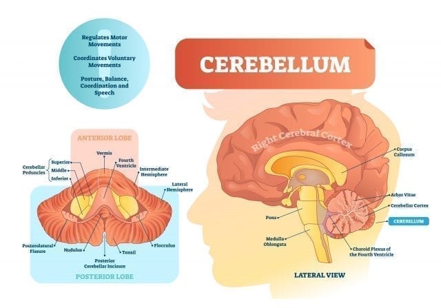
Cerebellum: Much more than motor coordination
It is likely that a few seconds ago before you arrived here you were typing on your computer or phone. We do it quickly and automatically, but… have you ever wondered how precise and harmonious the movement of your fingers is when you type? To get you to write correctly and efficiently, various structures are activated in our brain. The part of the brain in charge of coordinating these movements is the Cerebellum and it participates in many of the activities that we do every day: from walking to organizing a sentence.
What is the Cerebellum?
The Cerebellum is a brain structure partially concealed by the cortex. Classically it was thought that it was only in charge of harmonizing body movements, but for some years now it has become evident that it is involved in various cognitive functions. The Cerebellum has a shape similar to that of the brain, although a much smaller size. In fact, his name means “little brain.” It is divided into two hemispheres, and the portion of the cerebellum between them is called vermis. It is also the only part of the brain that has Purkinje Cells, a type of neurons essential for its functioning that allows the integration of the information it receives.
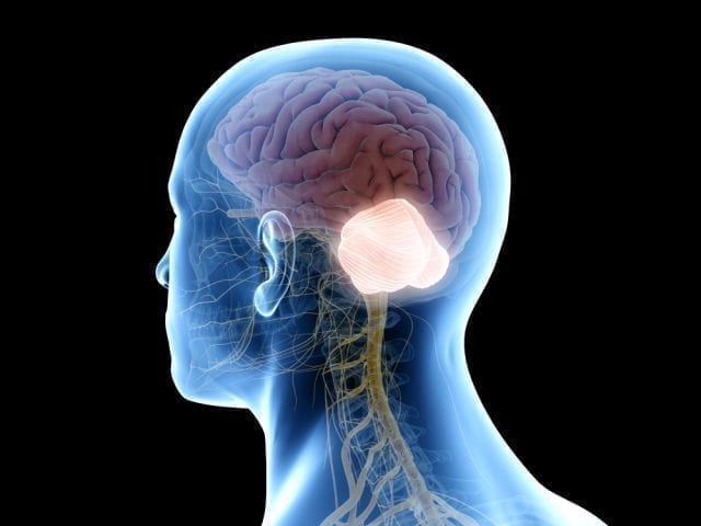
Where is the Cerebellum located? What parts does it consist of?
The Cerebellum is located in the back of the brain at the level of the brainstem bridge, under the occipital lobe (slightly above the nape of the neck). It binds to the rest of the brain through the lower, middle and upper cerebral peduncles, which are a set of nerve fibers that carry information from the rest of the body to the Cerebellum (afferent), or from the Cerebellum to the rest of the body (efferent). In fact, if it weren’t for the cerebral peduncles, it would be separated from the rest of the brain.
What is the Cerebellum for? Definition
The precision, harmony, and beauty of ballet dancers’ movements require a lot of dedication, practice and, above all, cerebellum. Each step of the choreography has a very determined force, rhythm, amplitude and, without the help of the cerebellum, all movement would be reduced to a set of spasms and falls (something that few people would be willing to pay for). However, in addition to this very important function of motor coordination, the cerebellum also participates in cognitive functions, without which ballet professionals could not, for example, reproduce movements by heart. Thus, the functions of the cerebellum are divided into motor functions and cognitive functions.
- Motor Functions of the Cerebellum: This structure receives information about, among other things, our equilibrium, the position of our body, which muscles we must move to perform a specific action, the direction of that movement and integrates it (gathers and works all of the above). When he has developed the information (something he does very, very quickly), he tells the rest of the brain how to carry out the movement. Thus, it regulates the intensity, speed, precise direction, travel and other characteristics of the movement so that, as a final result, we make a harmonious, precise and coordinated movement. To perform this function, the different parts of the cerebellum specialize in specific body parts, following a correspondence between the muscles and the surface of the cerebellum. A topographical representation has been made with this correspondence, called “homunculus of the Cerebellum”, which indicates which parts of the Cerebellum are in charge of which parts of the body.
- Cognitive Functions of the Cerebellum: For a relatively short time we have begun to study in depth what cognitive and emotional functions the cerebellum participates in. The most common way to investigate the functions of brain structures is to study the cases of people who have suffered some brain damage and, consequently, their cognitive abilities have been altered. Thus, more or less accurate conclusions can be drawn from the areas involved in the various functions (if X brain area is damaged and the person stops speaking, it is understood that this brain area participates in the ability to speak). The problem is that brain damage (such as stroke or head injury) is usually quite extensive, with more than one area affected as a result. This makes it difficult to study, as it is not known whether loss of function comes from damage in the X or Y area. However, different studies have investigated cognitive functions as well as the most recent advances, which allows us to know that the cerebellum contributes to the following cognitive processes:
- Language and Cerebellum: Participates in syntactic composition and grammar in general, in the articulation (that is really a motor function of the phonatory apparatus muscles), in the hidden articulation (that is, when we speak for ourselves in an internal dialogue, without making any noise), in the generation of words, in oral comprehension and in the establishment of a semantic relationship between words.
- Visuospatial skills: Complex visuospatial tasks such as the construction or mental rotation of images.
- Memory and learning (motor and non-motor): The cerebellum, along with other brain structures (basal ganglia), plays a major role in procedural memory (cycling, driving, writing your name in pencil or reading on the mirror) and in learning motor skills, habits and behaviors. It is also related to habituation and awareness, and to classical and operative conditioning. It is also activated by learning motor sequences and learning complex sequences. In conjunction with other structures (supplementary motor area and frontal operculum), the cerebellum participates in verbal operative memory, although it is not clear whether in internal coordination, in error settings, or both. On the other hand, the cerebellum can also take part in spatial memory.
- Executive Functions and Cerebellum: Executive functions are intimately related to the dorsolateral prefrontal cortex. However, being such a complex series of cognitive functions, they require the participation of other brain structures, including the cerebellum. The functions in which the cerebellum participates (although there is not much consistency in some of them) are planning, cognitive flexibility, abstract reasoning, working memory, verbal fluency, and inhibition. Some studies suggest that the cerebellum may be active during decision making or coordination of two tasks at the same time, increasing speed and automating new movements.
- Attention and cerebellum: In selective attention activities or other more complex functions that require attention, such as calculus, the cerebellum is involved.
- Personality, emotions, and cerebellum: Some studies point to the role of the cerebellum in controlling and modulating emotions. It has also been linked to personality in regulating appropriate or inappropriate behaviors.
Do you want to know the state of your cognitive processes?
There are currently clinical tools for this. The program that specialists recommend is CogniFit, the leading tool in neuropsychological assessments, cognitive training and stimulation tools. This brain stimulation program is based on cognitive reserve and neural plasticity to measure and improve mental performance through online games. The activities presented in this tool combine different neuropsychological batteries, therapeutic exercises, rehabilitation techniques and learning oriented to retrain and improve the skills that each person needs most.
This program can be used by health professionals, researchers, teachers and teaching staff, families, etc. It is a very easy to use the tool, so it is available to anyone who wants to know and improve their cognitive status.
CogniFit’s cognitive assessment and training tools enable you to measure, activate, exercise, and strengthen important cognitive abilities (attention, memory, executive functions, planning, perception, etc.) and the more than 20 components that comprise them.
All CogniFit cognitive stimulation programs have been validated so that children, adolescents, adults, and seniors can evaluate, activate and strengthen their mental capacity and compare their cognitive state with the rest of the world’s population.

Damage to the cerebellum does not paralyze any muscle, but it has important consequences. Some of these are:
- Ataxia: Ataxia is probably the most characteristic disorder derived from the alteration of the cerebellum. It consists of a movement disorder due to the inability to properly coordinate the different parts of the body involved. Errors of amplitude, speed, direction or force occur involuntary motor movements. Patients try to compensate for these errors, making coarse movements. Cerebellar ataxic gait is easily recognizable by uncoordinated and unbalanced walking. The problem is especially evident if the patient tries to walk with his or her eyes closed.
- Cerebellar dysplasia: Characterized by a scandalized or explosive speech (speaks in a jerky manner, with different intensities, in a disharmonic manner).
- Cerebellar nystagmus: It is an erratic, rapid and involuntary movement of the eyes.
- Dysmetria: This is the inability to properly coordinate the movement of your limbs with the visual information you receive. If you try to touch your nose, you miss since the moment has passed.
- Asynergy: The movements performed are carried out in a nonsynergistic way, that is, without coordination or harmony. The person tends to lose balance and adopt strange postures to compensate for this loss of balance.
- Adiadochokinesia: The inability to predict the positions of body parts when movement is performed.
- Intentional tremor: The tremor that occurs when a movement is made. Conversely, people with damage to the cerebellum do not usually have tremors at rest (being still).
- Hypotonia: The muscles are flaccid, as they have a lower tone than normal. Because of this and lack of balance, patients with cerebellar damage tend to perform many limb movements. Coordination tests show rebound phenomenon.
- Cognitive-affective cerebellar syndrome: When the cerebellum is affected, cognitive abilities and the control of related emotions are also altered, causing “inordinate thinking”. Cognitive abilities such as executive functions, attention, visual-spatial abilities, memory, language, or personality may undergo minor or severe changes.
Now that we know this, it is time to thank our cerebellum not only that we are able to walk, talk, type or dance in such a coordinated way, but also that it allows us to learn, structure the language correctly and plan our behaviors. In short, thank you for making it possible for us to live our daily lives with normality and harmony.
Anatomy: What are the cerebellum parts?
The cerebellum is a relatively large structure with a surface full of transverse grooves. According to these furrows, the Cerebellum is divided into the following lobes:
- Anterior lobe (Spinocerebellum or Paleocerebellum): It connects to the spinal cord. It is in charge of muscle tone, trunk, and limb movement.
- Posterior lobe (Brain cerebellum, Pontocerebellum or Neocerebellum): This is the portion of the cerebellum that is located near the Posterolateral Fissure. It takes care of voluntary movements and cognitive functions.
- Flocculonodular lobe (Vestibulocerebellum or Archicerebellum): It is the portion of the cerebellum below the Posterolateral Gap. Connects with vestibular and reticular nuclei. It takes care of balance, body position, head displacement and eye movements.
What nuclei is the Cerebellum composed of? What are they for?
The nuclei are a set of neural bodies that work in a coordinated way to carry out a series of more or less specific functions. The most important nuclei are:
- Nucleus fastigium (or of the roof). It receives the projections from the bark of the vermis.
- Globose nucleus (back interposition). The cortex that remains between the vermis and the two cerebellar hemispheres (paravermis) is projected into this nucleus.
- Emboliform nucleus. This nucleus also receives projections of the paravermis crust.
- Dentate nuclei: It is divided into three parts. It receives afferent, or incoming, signals from the premotor cortex and supplementary motor cortex via the pontocerebellar system
Cerebellar connections
In order to perform all its functions correctly, the cerebellum establishes a large number of input and output connections with various areas of the nervous system. However, it is an “isolated” structure from the rest. The only gateway to and from the information is the cerebellar peduncles. The peduncles are a set of afferent and efferent fibers that, depending on their position, can be divided into three pairs:
- Lower Cerebellar Peduncle: A set of fascicles that connect the spinal cord to the cerebellum and vice versa. It is mainly made up of afferent and some efferent fibers.
- Middle cerebellar peduncle: A set of fascicles that connect the brainstem bridge with the cerebellum and vice versa. It consists almost exclusively of afferent fibers.
- Upper cerebellar peduncle: A set of fascicles linking the midbrain to the cerebellum and vice versa. It is mainly made up of efferent fibers and some afferents.
In this way, the different connections of the cerebellum enter or exit through one or more of each pair of peduncles. If we look at what direction the information is going (if they enter or leave the Cerebellum), we distinguish between afferent and efferent, respectively. The afferent carry information from different parts of the body to the cerebellum. The main fascicles or tracts of afferent fibers are:
- Vestibulocerebellar fascicule: vestibular system – PCI – flocculonodular lobe.
- Spinocerebellar dorsal fascicule: Spinal cord – PCI – anterior lobe.
- Spinocerebellar ventral fascicule: Spinal cord -PCI and PCS -anterior lobe.
- Cuneocerebellar fascicule: Spinal bulb- PCI -anterior lobe.
- Olivocerebellar fascicle: Spinal bulb- PCI- anterior lobe.
- Reticulocerebellar fascicule: PCI and PCM-spinal lobe.
- Tectocerebellar fascicule: Mesencephalon – PCS – anterior lobe.
- Trigeminocerebellar fascicle: Mesencephalon – PCI and PCS -anterior lobe.
- Rubrocerebellar fascicle: Mesencephalon – PCS – anterior lobe.
- Corticoponticocerebellar fascicle: Cerebral cortex – PCM- posterior lobe.
On the other hand, the efferent refer to fibers that come out of the cerebellum and send information to other parts of the brain. The main efferent is:
- Cerebelovestibular fascicule: flocculonodular lobe- PCI- vestibular system.
- Motor flocculo-occulomotor fascicle: flocculonodular lobe -PCS – motor-occulo-vein.
- Cooked fascicule: flocculonodular lobe -PCI -vestibular system and oculomotor cores.
- Intermediateolivary fascicle: anterior lobe – PCS- the inferior olive core of the spinal bulb.
- Interproposedorreticular fascicle: anterior lobe – PCI – reticular formation.
- Interproposedorubic fascicle: anterior lobe – PCS – Red nucleus – Cerebral cortex.
- Interpostectal fascicule: anterior lobe – PCS -quadrigeminal tubers.
- Dentadotalamic fascicle: posterior lobe- PCS – Thalamus.
This article is originally written in Spanish by David Asensio, translated by Alejandra Salazar.